02 دی 1403
logo

مرکز تحقیقات استئوپروز

دانشگاه علوم پزشکی تهران

  • تاریخ انتشار : 1402/04/13 - 13:55
  • تعداد بازدید : 88
  • زمان مطالعه : 5 دقیقه

A Multiplexed Microfluidic Platform For Bone Marker Measurement: A Proof-Of-Concept

In this work, we report a microfluidic platform that can be easily translated into a biomarker diagnostic. This platform integrates microfluidic technology with electrochemical sensing and embodies a reaction/detection chamber to measure serum levels of different biomarkers. Microfabricated Au electrodes encased in a microfluidic chamber are functionalized to immobilize the antibodies, which can selectively capture the corresponding antigen.
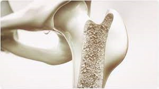
bone turnover markers {faces}
This is while enzyme-linked immunosorbent assay (ELISA), electrochemiluminescence immunoassay (ECLIA), and other prevailing laboratory techniques in the current state of the art for biomarker quantification do not lend themselves well to miniaturization for application at the point-of-care . Thus, performing rapid measurements to monitor several biochemical parameters at the same time to reach an accurate medical decision still remains a largely unmet challenge.
In this context, multiplexed detection of different biomarkers using microfluidic systems has attracted considerable interest . This is mainly due to the fact that such systems provide a promising method to miniaturize immunoassays, which have played an important role in the diagnosis of different diseases in the past 50 years. Such devices also provide added benefits, such as portability, increased reliability, improved sensitivity, decreased analysis time, minimal reagent consumption, and parallel processing . In other words, the high surface-to-volume ratio of the microfluidic channels facilitates faster and more sensitive reactions, which is of great importance in meeting the major demands for the on-site detection of various analytes .
Among different techniques used to generate signals in these platforms, electrochemical methods have shown the most suitable results because of their simple instrumentation, easy signal quantification, rapid response, low cost, portability, reliability, good sensitivity, and excellent selectivity . The sensitivity of biosensor platforms depend on various parameters, such as the microfluidic flow, as well as the specific surface area, conductivity, and charge transfer properties of the sensing platform . In this regard, functionalized gold nanoparticles (AuNPs) have shown promising results in improving the performance of the system through facilitating the electron exchange between the antibody complex and the electrode .
Despite the recent improvements in the development of multiplexed microfluidic-based systems, especially in personalized cancer diagnostics, a clear challenge still resides in the integration and operation of such microfluidic platforms for the measurement of different biomarkers in clinical practice . As such, a few attempts have been made in the past for the development of new screening assay methods for bone turnover markers (BTMs); yet none have succeeded to fully satisfy the criteria needed for their application in clinical practice . BTMs are important determinants of bone strength as they demonstrate the bone-remodeling rate by assessing bone resorption and formation . Monitoring the efficacy of bone-active drugs is currently the most promising clinical application for BTMs since, by using this technique, the changes in BTM levels in response to therapeutic interventions would be measurable in a shorter time interval (as early as 4–12 weeks) . Moreover, the pre-treatment levels are also useful in identifying the patients who will most benefit from the treatment. This is while “the routine use of biochemical markers of bone turnover in clinical practice is not generally recommended, as these tests vary in sensitivity and specificity and their appropriate role in patient management is not yet known . Thus, measuring bone mineral density (BMD) using dual energy X-ray absorptiometry (DXA) is currently considered as the gold standard for osteoporosis diagnosis despite its proven setbacks .
In the present work, we report the design and fabrication of a sensitive microfluidic platform integrated with electrochemical sensing that can easily be translated into a protein biomarker diagnostic. To our knowledge, this is the first microfluidic-based device designed as a step toward developing a PoC system for the measurement of several BTMs.

 Results

2.1. Osteokit Characterization

Having a well-dispersed layer of AuNPs with a uniform size distribution is the first and a critical step in surface modification. The deposition time and scan rate are, therefore, properly controlled to assure sufficient deposition and uniform distribution of AuNPs . The electrochemical behavior of the stepwise fabrication of the electrochemical sensor chip was studied using cyclic voltammetry (CV). Compared with conventional electrodes, the as-prepared AuNPs-based electrode-on-chip showed a 7.5-fold larger current response .
As mentioned in our previous article, the adsorption of the antibodies onto the working electrode was associated with a significant reduction of the conductive area of the working electrode, as well as a considerable increase in its electric resistance, resulting in a significant decrease in the magnitude of the electrochemical response current . This suggests successful immobilization of antibodies on the electrode.
Later, the presence of antigens on the working electrode resulted in an increase in the double-layer capacitance and, hence, reduced electrochemical behavior of the test solution proportional to the concentration of the antigen. This was used as the basis for the determination of antigen concentration and plotting the calibration curve.

2.2. Calibration of the Osteokit

The electrochemical response of the Osteokit was studied as a function of different concentrations of osteocalcin (Oc) and collagen type 1 cross-linked C-telopeptide (CTX) using chronoamperometry technique under identical experimental conditions (0.1 mM potassium ferricyanide [K3Fe(CN)6] as a redox probe, at a potential of 0.65 V for 15 s), respectively. It is noted that the sample consumption of this prototype chip was only 100 μL, which is less than traditional methods (10 mL).
The magnitude of response current ratio decreased with increasing antigen concentrations; this is due to the formation of immune complexes between the antibody and antigen, resulting in a hindrance in electron charge transfer rate at the electrode–electrolyte interface . The magnitude of current ratio in the Osteokit with a CTX immunoelectrode was linearly dependent on the logarithmic CTX concentration with an R2 of 0.78. The correlation coefficient was 0.99 and the limits of detection (LOD) and limits of quantification (LOQ) were calculated to be 1.39 pg/mL and 4.22 pg/mL, respectively.
  • Article_DOI : 10.3390/mi8050133
  • نویسندگان : patricia khashayar ,ghassem amoabediny,morteza hosseini,rik verplancke,farideh razi,jan vanfleteren,bagher larijani,david schaubroeck,steven van put,michel de keersmaecker,annemie adriaens,stefan goemaere,tom fiers
  • گروه خبر : مقالات,کارشناس مقالات
  • کد خبر : 237526
کبری گرگانی فیروزجاه
تهیه کننده:

کبری گرگانی فیروزجاه

0 نظر برای این مطلب وجود دارد

ارسال نظر

نظر خود را وارد نمایید:

متن درون تصویر را در جعبه متن زیر وارد نمائید *
متن مورد نظر خود را جستجو کنید
تنظیمات پس زمینه